This weekend saw a return visit to FSC Preston Montford for my final course in the University Certificate in Biological Recording and Species Identification – Identification of Macrofungi tutored by Irene Ridge. An interest in fungi (or mycology) was one of my earliest, after attending a fungi foray at Queen Elizabeth Country Park, Hampshire, with the RSPB’s Phoenix group way back in 1996. This led me to purchase a compound microscope in 2000 but I found it difficult to master the techniques needed to prepare slides without tuition and soon moved on to other groups, hence my motivation to attend this course and have another go.
After an introduction to fungi and how to record and describe specimens on the Friday evening it was off to Attingham Park on Saturday to collect our own specimens. ")

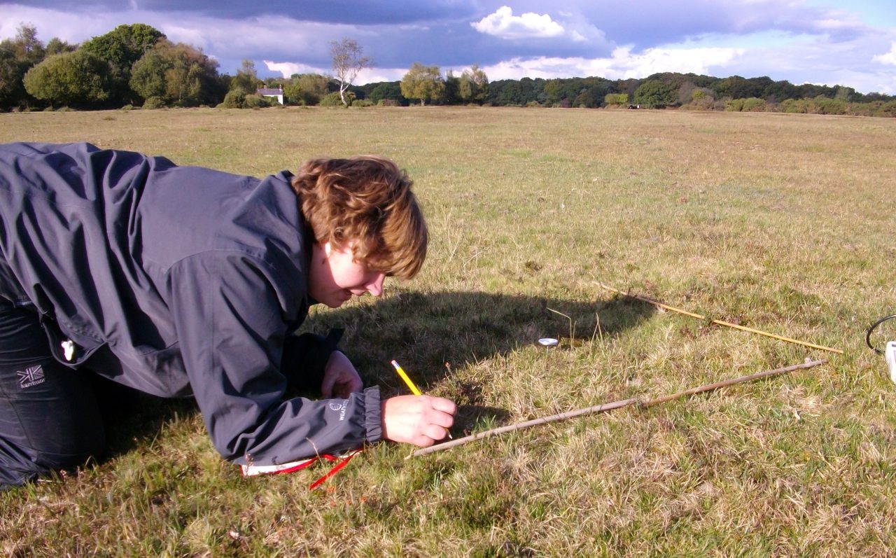
This site introduced me to grassland fungi, a habitat which I had not forayed before. The short, old lawns around Attingham Hall are an example of waxcap-grassland, a fungal community characterised by waxcaps (Hygrocybe spp.), club fungi (Clavariaceae) pink-gills (Entoloma spp.) and earthtongues (Geoglossaceae). Waxcap-grasslands are of conservation interest as indicators of unimproved grassland, a threatened habitat in the UK. It is only recently they have begun to receive attention on their ecology and distribution, with a English Nature Report published in 2003.
 |
|
Snowy Waxcap Hygrocybe virginea
|
 |
|
Apricot Club Clavulinopsis luteo-alba
|
Attingham is not all grassland though, there were many handsome mature trees in which we found some woodland fungi which were familiar to me. Irene explained the importance of taking good field notes when collecting fungi, many are mycorrhizal, a symbiotic relationship with plants, and some fungi species are restricted to certain tree species; without this information identification may not be possible. Whether the fungus was growing alone or in groups, was attached to wood or soil, and even the smell, colour and texture of the cap which may change after collection can be important for identification and must be noted on collection.
 on Beech at Attingham Park") |
| Artist’s Bracket Ganoderma sp. (probably G. applanatum) on Beech |
 |
|
A jelly-fungus, Exidea nucleolus
|
 |
| Irene discusses the identification features of a Boletus fungus |
Back in the lab we began identifying our collections using a mixture of identification books and keys. I began by making spore prints as the colour of the spores is a key feature in identification, in the past I had done this by cutting the cap off and putting it on white and black paper but Irene demonstrated suspending the fruit body over a microscope slide in a cup. This keeps the specimen moist, as spores do not drop when the atmosphere is dry, explaining why I was not very successful with mine in the past!
")
On the Sunday we were off on another trip, this time to woodland at Nesscliffe Country Park.
 |
|
Beech woodland at Nesscliffe – lots of lovely leaf litter!
|
 |
|
Russula ochroleuca
|
 |
|
Jelly Ear Auricularia auricula-judae
|
 at Nesscliffe") |
") |
Another old favourite is “smell it before you see it” Stinkhorn (Phallus impudicus) although we only found in the ‘egg’ stage and one old specimen, both badly eaten by slugs, missing out on in its erect magnificence.
 |
|
Stinkhorn (Phallus impudicus) ‘egg’
|
 |
| Basidia stained with Congo Red, the pointy bits on top are where the spores attach
By Andreas Kunze (Own work) [CC-BY-SA-3.0 (http://creativecommons.org/licenses/by-sa/3.0)], via Wikimedia Commons) |
 |
|
Cystidia
By Andreas Kunze (Own work) [CC-BY-SA-3.0 (http://creativecommons.org/licenses/by-sa/3.0)], via Wikimedia Commons)
|
The course finished with an assignment – identifying 15 specimens and/or photographs, it was strange to think it was my last on the course and that with my uCert and MSc complete I am for the first time in 7 years not in formal education! The two fungi forays and tuition in microscopy techniques were just what I needed to get back into fungi identification, I will be dusting off my old compound microscope and giving it another go!
